

1. What is skin cancer?
Skin cancer is the out-of-control growth of abnormal cells in the epidermis, the outermost skin layer, caused by unrepaired DNA damage that triggers mutations. These mutations lead the skin cells to multiply rapidly and form malignant tumors.

Picture: Skin Cancer
2. What are the main types of skin cancer?
Basal cell carcinoma
Squamous cell carcinoma
Skin adnexal tumors (e.g. sebaceous carcinoma)
Melanoma
Merkel cell carcinoma
Sarcomas of primary cutaneous origin (e.g. dermatofibrosarcoma protuberans)
Lymphomas of primary cutaneous origin (e.g. mycosis fungoides)
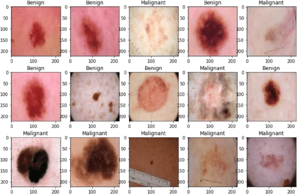
Picture: Diferent Types of Skin Cancer
4. How to prevent skin cancer?
Most skin cancers are preventable. To protect yourself, follow these skin cancer prevention tips:
· Avoid the sun during the middle of the day.
· Wear sunscreen year-round.
· Wear protective clothing
· Avoid tanning beds.
· Be aware of sun-sensitizing medications.
· Check your skin regularly and report changes to your doctor.
5. What is the most aggressive form of skin cancer?
Melanoma is considered the most dangerous form of skin cancer as it typically will spread to other areas of the body, including organs, if left untreated.
6. What is the most common skin cancer?
Basal cell carcinoma (also called basal cell skin cancer) is most common type of skin cancer. About 8 out of 10 skin cancers are basal cell carcinomas (also called basal cell cancers).
7. Can you die from skin cancer?
About 2,000 people die from basal cell and squamous cell skin cancer each year. Older adults and people with a suppressed immune system have a higher risk of dying from these types of skin cancer. About 7,180 people die from melanoma each year.
8. How do you know if a spot is skin cancer?
Redness or new swelling beyond the border of a mole. Color that spreads from the border of a spot into surrounding skin. Itching, pain, or tenderness in an area that doesn’t go away or goes away then comes back. Changes in the surface of a mole: oozing, scaliness, bleeding, or the appearance of a lump or bump.
9. What can be mistaken for skin cancer?
To help put things into perspective here are 5 skin conditions that are often mistaken for skin cancer:
· Psoriasis. …
· Seborrheic Keratoses (Benign tumour) …
· Sebaceous hyperplasia. …
· Nevus (mole) …
· Cherry angioma.
10. What Colour is skin cancer?
Pigmented basal cell cancers have dark areas, often brown, blue or grey in colour. They can look like warts or sometimes melanoma.
11. Are skin cancers itchy?
Yes, skin cancer can be itchy. For example, basal cell skin cancer can appear as a crusty sore that itches. The deadliest form of skin cancer — melanoma — can take the form of itchy moles. See your doctor for any itchy, crusty, scabbed, or bleeding sore that’s not healing.
12. How quickly does skin cancer spread?
Melanoma can grow very quickly. It can become life-threatening in as little as six weeks and, if untreated, it can spread to other parts of the body. Melanoma can appear on skin not normally exposed to the sun.
13. What cancers cause itching?
The cancers that are most commonly associated with itching are lymphoma, polycythaemia Vera (PV), certain gastrointestinal cancers, and melanoma.
14. Where is skin cancer most common?
Skin cancer develops primarily on areas of sun-exposed skin, including the scalp, face, lips, ears, neck, chest, arms and hands, and on the legs in women. But it can also form on areas that rarely see the light of day — your palms, beneath your fingernails or toenails, and your genital area.
15. Can skin cancer appear overnight?
It can appear suddenly, but they can also grow slowly over time. It’s most common in older individuals, especially those who have fair skin.
16. Does skin cancer grow slowly?
The cancer lesion often appears as small, raised, shiny, or pearly bumps, but it can have various kinds of appearance. They tend to grow slowly and rarely spread to other parts of the body.
17. What is Basal and Squamous Cell Skin Cancer
These cancers are most often found in areas exposed to the sun, such as the head, neck, and arms, but they also can occur elsewhere. They are very common but are also usually very treatable. Here you can find out all about these cancers, including risk factors, symptoms, how they are found, and how they are treated.
18. What are the types of basal cell carcinoma?
CLINICAL VARIANTS OF BASAL CELL CARCINOMA
· Nodular basal cell carcinoma
· Cystic BCC
· Sclerodermiform (Morpheiform) BCC.
· Infiltrated basal cell carcinoma.
· Micronodular basal cell carcinoma.
· Superficial basal cell carcinoma.
· Pigment basal cell carcinoma.
· Fibroepithelioma of Pinkus.
19. What are the different types of squamous cell carcinoma?
The primary types of squamous cell carcinoma are:
· Adenoid/pseudoglandular squamous cell carcinoma.
· Intraepidermal squamous cell carcinoma.
· Large cell keratinizing squamous cell carcinoma.
· Large cell non-keratinizing squamous cell carcinoma.
· Lymphoepithelial carcinoma.
· Papillary squamous cell carcinoma.
20. What is the difference between Bowen’s disease and squamous cell carcinoma?
Bowen’s disease is a skin condition that has continuous, slow-developing scaly-red patches. It is also known as squamous cell carcinoma in situ (SCC in situ), and is a non-invasive and presiding form of intraepidermal carcinoma. However, it has only a 10% chance of developing into SCC, a type of common skin cancer.
21. How can you tell the difference between squamous cell carcinoma and actinic keratosis?
The main difference between SCC in situ and AK is that in SCC in situ, the full thickness of the epidermis is involved with atypical proliferation of keratinocytes; whereas, in AK, the atypia is limited to lower levels of the epidermis and not its full thickness.
22. Is squamous cell carcinoma malignant or benign?
Benign skin cancers, such as squamous cell carcinoma (SCC), typically develop due to overexposure to the sun and appear on various parts of the body, such as the nose, forehead, lower lip, ears, and hands.
23. What is the best treatment for squamous cell carcinoma?
The simplest and most common treatment for smaller SCC in situ is surgical excision. The standard practice is to remove about a quarter inch beyond the edge of the cancer. Larger ones can also be excised, but Mohs surgery may be needed. It offers the highest cure rate of all treatment methods.
24. How fast does squamous cell carcinoma grow?
Results: Rapidly growing SCC occurred most commonly on the head and neck, followed by hands and extremities, and had an average duration of 7 weeks before diagnosis. The average size of the lesions was 1.29 cm and nearly 20% occurred in immunosuppressed patients.
25. Do you need chemo for squamous cell carcinoma?
Larger squamous cell cancers are harder to treat, and fast-growing cancers have a higher risk of coming back. In rare cases, squamous cell cancers can spread to lymph nodes or distant parts of the body. If this happens, treatments such as radiation therapy, immunotherapy, and/or chemotherapy may be needed.
26. What happens if squamous cell carcinoma spreads to lymph nodes?
When squamous cell cancer spreads to lymph nodes in the neck or around the collarbone, it is called metastatic squamous neck cancer. The doctor will try to find the primary tumor (the cancer that first formed in the body), because treatment for metastatic cancer is the same as treatment for the primary tumor.
27. Why does squamous cell carcinoma keep coming back?
That’s because individuals who were diagnosed and treated for a squamous cell skin lesion have an increased risk of developing a second lesion in the same location or a nearby skin area. Most recurrent lesions develop within two years after the completion of treatment to remove or destroy the initial cancer.
28. What organs does squamous cell carcinoma affect?
Squamous cell carcinoma can spread to other parts of the body, including fatty tissues, lymph nodes, and internal organs. It can cause death. Squamous cell carcinomas located on the lip, ears, palm of the hand, or sole of the foot have the highest risk of spreading.
29. What is the mortality rate of squamous cell carcinoma?
Squamous cell carcinoma (SCC) generally has a high survival rate. The 5-year survival is 99 percent when detected early. Once SCC has spread to the lymph nodes and beyond, the survival rates are lower. Yet this cancer is still treatable with surgery and other therapies, even in its advanced stages.
30. Does squamous cell carcinoma have roots?
Squamous cell skin cancer (Squamous Cell Carcinoma or SCC)
This form of skin cancer grows more quickly, and though it can be confined to the top layer of skin, it frequently grows roots. Squamous cell carcinoma can be more aggressive and does have a potential to spread internally.
31. Does squamous cell carcinoma appear suddenly?
It is a rapidly growing tumor which tends to appear suddenly and may reach a considerable size. This tumor is often dome-shaped with a central area resembling a crater which is filled with a keratin plug.
32. How long does it take to recover from squamous cell carcinoma surgery?
Depending upon the size, may take up to 4 to 6 weeks for the wound to heal completely, but infection, bleeding and pain are uncommon. Close the wound with sutures (stitches). This option is appropriate when scarring must be kept to a minimum or when the natural healing process would be inadequate.
33. What are Basal and Squamous Cell Skin Cancer Risk Factors?
Several risk factors make a person more likely to get basal cell or squamous cell skin cancer. Ultraviolet (UV) light exposure, Having light-colored skin/ albinism, Being older, Being male, Exposure to certain chemicals, Radiation exposure, Previous skin cancer, Long-term or severe skin inflammation or injury, Psoriasis treatment, Xeroderma pigmentosum (XP), Basal cell nevus syndrome (also known as nevoid basal cell carcinoma syndrome or Gorlin syndrome), Weakened immune system, Human papillomavirus (HPV) infection,Smoking.
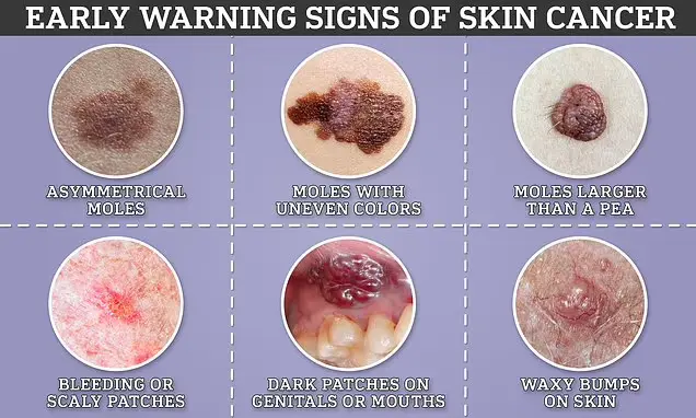
Picture: Signs of Skin Cancer
34. Can Basal and Squamous Cell Skin Cancers Be Prevented?
There is no sure way to prevent all basal and squamous cell skin cancers. Some risk factors such as your age, gender, race, and family history can’t be controlled. But there are things you can do that could lower your risk of getting these and other skin cancers.
35. What are sign and symptoms of basal cell cancer?
Individuals with a basal-cell carcinoma typically present with a shiny, pearly skin nodule. However, superficial basal-cell cancer can present as a red patch similar to eczema. Infiltrative or morpheaform basal-cell cancers can present as a skin thickening or scar tissue – making diagnosis difficult without using tactile sensation and a skin biopsy. It is often difficult to visually distinguish basal-cell cancer from acne scar, actinic elastosis, and recent cryodestruction inflammation
36. Where do basal cell carcinoma develop?
These cancers usually develop on sun-exposed areas, especially the face, head, and neck. They tend to grow slowly. It’s very rare for a basal cell cancer to spread to other parts of the body.
37. What happens if basal cell cancer left untreated?
if it’s left untreated, basal cell cancer can grow into nearby areas and invade the bone or other tissues beneath the skin.
38. Is a basal cell carcinoma dangerous?
How dangerous is BCC? While BCCs rarely spread beyond the original tumor site, if allowed to grow, these lesions can be disfiguring and dangerous. Untreated BCCs can become locally invasive, grow wide and deep into the skin and destroy skin, tissue and bone.
39. Is basal cell carcinoma malignant or benign?
Basal cell carcinoma (BCC) is most often a benign form of skin cancer caused by exposure to ultraviolet (UV) light. However, it’s the most frequently occurring form of all skin cancers.
40. Can basal cell carcinoma turn into melanoma?
Basal cell carcinoma does not progress into melanoma. Each is a separate and distinct type of skin cancer. Basal cell carcinoma is the most common form of skin cancer and one of two major nonmelanoma skin cancer types (the other is squamous cell carcinoma).
41. What is worse squamous or basal cell carcinoma?
Though not as common as basal cell (about one million new cases a year), squamous cell is more serious because it is likely to spread (metastasize). Treated early, the cure rate is over 90%, but metastases occur in 1%–5% of cases. After it has metastasized, it’s very difficult to treat.
42. Where do skin cancers start?
Most skin cancers start in the top layer of skin, called the epidermis. There are 3 main types of cells in this layer:
- Squamous cells: These are flat cells in the upper (outer) part of the epidermis, which are constantly shed as new ones form. When these cells grow out of control, they can develop into squamous cell skin cancer (also called squamous cell carcinoma).
- Basal cells: These cells are in the lower part of the epidermis, called the basal cell layer. Skin cancers that start in the basal cell layer are called basal cell skin cancers or basal cell carcinomas.
- Melanocytes: Melanoma skin cancer starts in these cells.
The epidermis is separated from the deeper layers of skin by the basement membrane. When a skin cancer becomes more advanced, it generally grows through this barrier and into the deeper layers.
43. How quickly does basal cell carcinoma spread?
The tumors enlarge very slowly, sometimes so slowly that they go unnoticed as new growths. However, the growth rate varies greatly from tumor to tumor, with some growing as much as ½ inch (about 1 centimeter) in a year.
44. What is considered a large basal cell carcinoma?
A size larger than 3 cm has been described as a high-risk feature. Notwithstanding the foregoing, this risk factor has been more accurately defined as 1 cm for head and neck tumors and more than 2 cm in other body areas.
45. How do they cut out basal cell carcinoma?
1. Surgical excision. In this procedure, your doctor cuts out the cancerous lesion and a surrounding margin of healthy skin. …
2. Mohs surgery. During Mohs surgery, your doctor removes the cancer layer by layer, examining each layer under the microscope until no abnormal cells remain.
46. What is invasive basal cell carcinoma?
Basal cell carcinoma (BCC) is a common, locally invasive, keratinocyte cancer (also known as nonmelanoma cancer). It is the most common form of skin cancer. BCC is also known as rodent ulcer and basalioma. Patients with BCC often develop multiple primary tumours over time.
47. Is Basal Cell Carcinoma genetic?
Basal cell carcinoma (BCC) and squamous cell carcinoma (SCC) are two of the most common malignancies in the United States and are often caused by sun exposure, although several hereditary syndromes and genes are also associated with an increased risk of developing these cancers.
48. Can basal cell carcinoma be frozen off?
Cryotherapy is a nonsurgical treatment for basal cell carcinoma. Your doctor applies liquid nitrogen to the tumor, freezing the abnormal tissue. The frozen skin then sloughs off (falls away) as the skin underneath heals.
49. Is nodular basal cell carcinoma invasive?
Micronodular BCCs contain small nodules of tumor cells that invade surrounding stroma. The morpheaform and micronodular types are generally the most locally invasive variants of BCC.
50. Can basal cell carcinoma shrink on its own?
In very rare cases, then, a basal cell carcinoma tumor can shrink and disappear on its own without treatment. Unfortunately, that doesn’t mean the cancer has gone away. There is an increased risk the cancer will return or reactivate. Most tumors, however, will grow larger over time and can cause significant damage.
51. Does basal cell carcinoma have roots?
BCCs have roots around and below the visible lesion. The roots can only be seen with a microscope. The lesion enlarges as the roots expand, similar to a weed. If the roots are not treated, then the BCC will come back – just like a weed.
52. How do you get rid of basal cell carcinoma without surgery?
Basal Cell Skin Cancer Treatment
Patients now have another option: Superficial Radiation Therapy, or SRT — a non-surgical treatment for basal cell carcinoma. It is the non-surgical choice for removing non-melanoma skin cancer.
53. Is Mohs surgery necessary for basal cell carcinoma?
Patients should also consider Mohs surgery when a BCC has recurred or has an aggressive growth pattern or poorly defined borders. Mohs surgery is a first-line choice for many BCC patients.
54. Can patient go in the sun after basal cell carcinoma?
If you’ve had skin cancer, you should avoid spending too long in the sun. Use a sunscreen product with at least a sun protector factor (SPF) of 15 and apply generously. Your skin cancer specialist might suggest a high factor sunscreen such as 50 on any exposed skin.
55. What is melanoma?
Melanoma, also redundantly known as malignant melanoma is a type of skin cancer that develops from the pigment-producing cells known as melanocytes.
56. What are signs and symptoms of melanoma?
Early signs of melanoma are changes to the shape or color of existing moles or, in the case of nodular melanoma, the appearance of a new lump anywhere on the skin. At later stages, the mole may itch, ulcerate or bleed. Early signs of melanoma are summarized by the mnemonic “ABCDEF”
· Asymmetry
· Borders (irregular with edges and corners)
· Colour (variegated)
· Diameter (greater than 6 mm (0.24 in), about the size of a pencil eraser)
· Evolving over time
· Elevated above the skin surface
· Firm to the touch
· Growing
Metastatic melanoma may cause nonspecific paraneoplastic symptoms, including loss of appetite, nausea, vomiting and fatigue. Metastasis (spread) of early melanoma is possible, but relatively rare: less than a fifth of melanomas diagnosed early become metastatic. Brain metastases are particularly common in patients with metastatic melanoma. It can also spread to the liver, bones, abdomen or distant lymph nodes.
57. What is the primary cause of melanoma?
The primary cause of melanoma is ultraviolet light (UV) exposure in those with low levels of the skin pigment melanin. The UV light may be from the sun or other sources, such as tanning devices. Those with many moles, a history of affected family members and poor immune function are at greater risk. A number of rare genetic conditions such as xeroderma pigmentosum also increase the risk.
58. How melanoma is diagnosed?
Diagnosis is by biopsy and analysis of any skin lesion that has signs of being potentially cancerous. To detect melanomas (and increase survival rates), it is recommended to learn to recognize them to regularly examine moles for changes (shape, size, color, itching or bleeding) and to consult a qualified physician when a candidate appears. dermoscopy is more helpful to identify malignant lesions than use of the naked eye alone. Reflectance confocal microscopy may have better sensitivity and specificity than dermoscopy in diagnosing cutaneous melanoma.
59. What are the Histopathologic types of melanoma?
These are types of melanoma.
· Superficial spreading melanoma
· Nodular melanoma
· Lentigo maligna melanoma
· Acral lentiginous melanoma
· Mucosal melanoma; When melanoma occurs on mucous membranes.
· Desmoplastic melanoma
· Melanoma with small nevus-like cells
· Melanoma with features of a Spitz nevus
· Uveal melanoma
· Vaginal melanoma
· Polypoid melanoma, a subclass of nodular melanoma.
60. Is melanoma invasive?
Some histopathological types of melanoma are inherently invasive, including nodular melanoma and lentigo maligna melanoma, where the in situ counterpart to lentigo maligna melanoma is lentigo maligna. Lentigo maligna is sometimes classified as a very early melanoma and sometimes a precursor to melanoma.
Superficial spreading melanomas and acral lentiginous melanomas can be either in situ or invasive, but acral lentiginous melanomas are almost always invasive.
61. What are the factors that affect melanoma prognosis?
Factors that affect prognosis include:
· tumor thickness in milli meters (Breslow’s depth),
· depth related to skin structures (Clark level),
· type of melanoma,
· presence of ulceration,
· presence of lymphatic/ perineural invasion,
· presence of tumor-infiltrating lymphocytes (if present, prognosis is better),
· location of lesion,
· presence of satellite lesions, and
· presence of regional or distant metastasis.
62. How long does it take a Melanoma to spread?
Melanoma can grow very quickly. It can become life-threatening in as little as six weeks and, if untreated, it can spread to other parts of the body.
63. What is the most common treatment for melanoma?
The main treatment for melanoma is surgical removal, or excision, of the primary melanoma on the skin. The extent of the surgery depends on the thickness of the melanoma. Most melanomas are found when they are less than 1.0 mm thick, and outpatient surgery is often the only treatment needed.
64. How stage 1 & 2 melanoma is treated?
Treatment of stage 1&2 melanoma may include the following:
· Surgery to remove the tumor and some of the normal tissue around it. Sometimes lymph node mapping and removal of lymph nodes is also done.
· A clinical trial of new ways to find cancer cells in the lymph nodes.
65. What is the treatment of Stage III Melanoma?
Treatment of stage III melanoma that can be removed by surgery may include the following:
· Surgery to remove the tumor and some of the normal tissue around it.
· Surgery followed by immunotherapy with immune checkpoint inhibitors (nivolumab, pembrolizumab, or ipilimumab) if there is a high risk that the cancer will come back.
· Surgery followed by targeted therapy with signal transduction inhibitors (dabrafenib and trametinib) if there is a high risk that the cancer will come back.
· A clinical trial of immunotherapy with or without vaccine therapy.
· A clinical trial of surgery followed by therapies that target specific gene changes.
66. How to treat Stage III Melanoma That Cannot Be Removed By Surgery, Stage IV Melanoma, and Recurrent Melanoma?
Treatment of stage III melanoma that cannot be removed by surgery, stage IV melanoma, and recurrent melanoma may include the following:
· Oncolytic virus therapy (talimogene laherparepvec) injected into the tumor.
· Immunotherapy with ipilimumab, pembrolizumab, nivolumab, or interleukin-2 (IL-2). Sometimes ipilimumab and nivolumab are given together.
· Targeted therapy with signal transduction inhibitors (dabrafenib, trametinib, vemurafenib, cobimetinib, encorafenib, binimetinib). These may be given alone or in combination.
· Chemotherapy.
· Palliative therapy to relieve symptoms and improve the quality of life. This may include:
o Surgery to remove lymph nodes or tumors in the lung, gastrointestinal (GI) tract, bone, or brain.
o Radiation therapy to the brain, spinal cord, or bone.
67. Which treatments are in clinical trials for Stage III Melanoma That Cannot Be Removed By Surgery, Stage IV Melanoma, and Recurrent Melanoma?
Treatments that are being studied in clinical trials include the following:
· Immunotherapy alone or in combination with other therapies such as targeted therapy.
· For melanoma that has spread to the brain, immunotherapy with nivolumab plus ipilimumab.
· Targeted therapy, such as signal transduction inhibitors, angiogenesis inhibitors, oncolytic virus therapy, or drugs that target certain gene mutations. These may be given alone or in combination.
· Surgery to remove all known cancer.
· Regional chemotherapy (hyperthermic isolated limb perfusion). Some patients may also have immunotherapy with tumor necrosis factor.
· Systemic chemotherapy.
68. What percentage of melanoma is fatal?
The estimated five-year survival rate for patients whose melanoma is detected early is about 99 percent. The survival rate falls to 66 percent when the disease reaches the lymph nodes and 27 percent when the disease metastasizes to distant organs.
69. What is Superficial spreading melanoma?
Superficial spreading melanoma (SSM) is usually characterized as the most common form of cutaneous melanoma in Caucasians. The average age at diagnosis is in the fifth decade, and it tends to occur on sun-exposed skin, especially on the backs of males and lower limbs of females.
70. What are signs and symptoms of SSM?
A prolonged radial growth phase, where the lesion remains thin, may eventually be followed by a vertical growth phase where the lesion becomes thick and nodular.
71. How SSM can be treated?
Treatment is by excisional biopsy, wide local excision and possibly sentinel node biopsy. Spread of disease to local lymph nodes or distant sites (typically brain, bone, skin and lung) marks a decidedly poor prognosis.
72. What is Nodular melanoma?
Nodular melanoma (NM) is the most aggressive form of melanoma. It tends to grow more rapidly in thickness (vertically penetrate the skin) than in diameter compared to other melanoma subtypes.
73. What are important prognosis factors for NM ?
Important prognosis factors for nodular melanoma include:
· Thickness
· Ulceration
· Sentinel lymph node (SLN) status
74. What are treatment plans for NM?
Therapies for metastatic melanoma include the biologic immunotherapy agents ipilimumab, pembrolizumab, and nivolumab; BRAF inhibitors, such as vemurafenib and dabrafenib; and a MEK inhibitor trametinib.
75. What is Lentigo maligna melanoma
Lentigo maligna melanoma is a melanoma that has evolved from a lentigo maligna, as seen as a lentigo maligna with melanoma cells invading below the boundaries of the epidermis.
76. Where LMM can be found?
They are usually found on chronically sun damaged skin such as the face and the forearms of the elderly.
77. Is LMM invasive?
Lentigo maligna is the non-invasive skin growth that some pathologists consider to be a melanoma-in-situ. A few pathologists do not consider lentigo maligna to be a melanoma at all, but a precursor to melanomas. Once a lentigo maligna becomes a lentigo maligna melanoma, it is treated as if it were an invasive melanoma.
78. Where ALM can be found?
Acral lentiginous melanoma is observed on the palms, soles, under the nails and in the oral mucosa. It occurs on non-hair-bearing surfaces of the body, which have not necessarily been exposed to sunlight. It is also found on mucous membranes.
79. What causes ALM’s?
Acral lentiginous melanoma is a result of malignant melanocytes at the membrane of the skin (outer layers).The pathogenesis of acral lentiginous melanoma remains unknown at this time.
80. What are histological signs of ALM?
The main characteristic of acral lentiginous melanoma is continuous proliferation of atypical melanocytes at the dermoepidermal junction. Other histological signs of acral lentiginous melanoma include dermal invasion and desmoplasia.
81. How ALMs are treated?
Therapies for metastatic melanoma include the biologic immunotherapy agents ipilimumab, pembrolizumab, and nivolumab; BRAF inhibitors, such as vemurafenib and dabrafenib; and a MEK inhibitor trametinib.
82. What is Desmoplastic melanoma?
Desmoplastic melanoma is a rare cutaneous condition characterized by a deeply infiltrating type of melanoma with an abundance of fibrous matrix.It usually occurs in the head and neck region of older people with sun-damaged skin.
83. What is uveal melanoma?
Uveal melanoma is a cancer (melanoma) of the eye involving the iris, ciliary body, or choroid (collectively referred to as the uvea). Tumors arise from the pigment cells (melanocytes) that reside within the uvea and give color to the eye. These melanocytes are distinct from the retinal pigment epithelium cells underlying the retina that do not form melanomas. When eye melanoma is spread to distant parts of the body, the five-year survival rate is about 15%.
84. What are the types of melanoma?
Uveal melanomas, often referred to by the media and in the general population as ocular melanomas, may arise from any of the three parts of the uvea, and are sometimes referred to by their location, choroidal melanoma, ciliary body melanoma, or iris melanoma.
85. What are the signs and symptoms of Uveal melanoma?
When symptoms do occur, they can include:
· blurred vision
· double vision (diplopia)
· irritation
· pain
· a perception of flashes of light in the eye (photopsia)
· a reduction in the total field of vision
· loss of vision
· a sensation of a foreign body in the field of vision (floaters)
· redness, bulging or displacement of the eye (proptosis),
· a change in the shape of the pupil
· pressure within the eye
· metamorphopsia (a distortion of vision where, when a person looks at a grid of straight lines, the lines appear wavy and parts of the grid appears blank).
86. How uveal melanoma can be diagnosed?
It can be diagnosed by using following methods:
· clinical examination by biomicroscopy
· indirect ophthalmoscopy
87. What is the treatment method used for UM?
Following treatment methods are considered:
· Brachytherapy,
· Enucleation
· Proton beam radiotherapy
· Transpupillary thermotherapy
· Photocoagulation
· Photodynamic therapy
· Local resection.
88. How UM can be prevented?
It can be prevented by reducing exposure to UV light.
89. What are the causes of uveal melanoma?
The cause of uveal melanoma is unclear. Uveal nevi are common (5% of Caucasians),but rarely progress to melanoma.
90. Is uveal cancer metastatic?
There are no lymphatic channels to the uveal tract, metastasis occurs through local extension and/or blood-borne dissemination. The most common site of metastasis for uveal melanoma is the liver, the liver is the first site of metastasis for 80%-90% of ocular melanoma patients. Other common sites of metastasis include the lung, bones, and just beneath the skin (subcutaneous).
91. What are the factors affecting the etiology of UM metastasis?
Several clinical and pathological prognostic factors have been identified that are associated with higher risk of metastasis of uveal melanomas. These include large tumor size, ciliary body involvement, presence of orange pigment overlying the tumor, and older patient age. Likewise several histological and cytological factors are associated with higher risk of metastasis, including presence and extent of cells with epithelioid morphology, presence of looping extracellular matrix patterns, increased infiltration of immune cells, and staining with several immunohistochemical markers.
92. What is the role of BAP1 in poorprognosis of uveal cancer?
The most important genetic alteration associated with poor prognosis in uveal melanoma is inactivation of BAP1, which most often occurs through mutation of one allele and subsequent loss of an entire copy of chromosome 3 (monosomy 3) to unmask the mutant copy. Because of this function in inactivation of BAP1, monosomy 3 correlates strongly with metastatic spread.
93. What is the most accurate prognostic factor for uveal cancer?
The most accurate prognostic factor is molecular classification by gene expression profiling of uveal melanomas. This analysis has been used to identify two subclasses of uveal melanomas: class 1 tumors that have a very low risk of metastasis, and class 2 tumors that have a very high risk of metastasis. Gene expression profiling outperforms all factors at predicting metastatic spread of the primary tumor.
94. What is occurrence rate of uveal cancer?
Uveal melanomas are the most common primary intraocular tumor in adults. Uveal melanoma is classified as a rare cancer with 5.1 cases per million people per year.The incidence has remained stable for several years.
95. What is the survival rate of uveal melanoma?
When eye melanoma is spread to distant parts of the body, the five-year survival rate is about 15%.
96. What is vaginal melanoma?
Vaginal melanoma is a rare malignancy that originates from melanocytes in the vaginal epithelium. It is also known as a melanocytic tumor or as a malignant melanoma. It is aggressive and infrequently cured. The median overall survival is 16 months. Vaginal melanoma accounts 5.5% of all vaginal cancers and only 1% of all melanomas diagnosed in women. Vaginal melanomas are frequently diagnosed in advanced stages of the disease. The prognosis is poor and the most important risk factor is the presence of lymph node metastases.
➰ Visit your trusted source: MAYO CLINIC